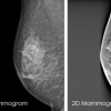


Stereotactic biopsy technique is the preferred way to biopsy microcalcifications (a tiny cluster of small calcium deposits) detected on mammography and it can be used to biopsy non-palpable nodules which are not visible on ultrasound.
Afterwards
When the biopsies have been taken, the radiographer will image the specimen to confirm that there are calcifications present. Once confirmed, the compression will be released and a dressing is applied. This can be removed after two days. The specimen is sent to the laboratory for analysis. Results are usually available within 5-7 working days. You will see one of our breast surgeons for the results.

How does Stereotactic biopsy work?
We use digital mammography plus computer aided geometry to accurately localise the area of concern within the breast. This allows the radiologist to place a biopsy needle in the correct position following infiltration of local anaesthetic and to obtain biopsy samples.

The Procedure
You will be positioned either sitting or lying close to the mammogram machine with your breast compressed. Some check images will be obtained to localise the area of concern. Then you will be given local anaesthetic into the skin. The biopsy needle is then advanced to the lesion and the samples taken. The procedure is not usually painful, but some women find it uncomfortable as there is prolonged breast compression.
We advise you not to do anything strenuous for the remainder of the day. You can take paracetamol every four hours for any pain and discomfort.